Tracking residual tumor after surgery is essential for catching recurrence early, but automating post-operative glioma segmentation remains a difficult task. Although transformer-based architectures, such as SwinUNETR, achieved impressive results, few studies test how well they generalize across clinical protocols.
In this paper, we conduct an ablation study on the MU-GLIOMA-POST and UCSF-ALPTDG datasets and show that the standard Generalized Dice Loss (GDL) is unstable under domain shift: the Whole Lesion (WL) Dice drops from 0.88 on the internal validation set to 0.73 on the external UCSF test set. To address this, we pair brain-masked percentile normalization with voxel-level contrastive learning.
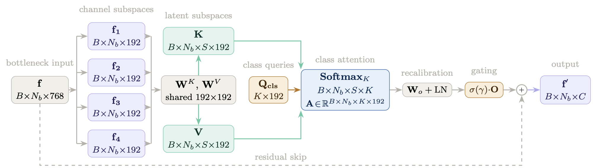
We also propose a Subspace-Aware Class Attention (SACA) module that re-calibrates the bottleneck features and raises Enhancing Tumor (ET) sensitivity by 8% (9.1% relative improvement) on internal validation. Ensembling these refinements with nnU-Net brings every stable configuration to a WL Dice of 0.94, and the SACA variant ensemble achieves the best boundary error (HD95) of 2.92 mm on MU-GLIOMA-POST.
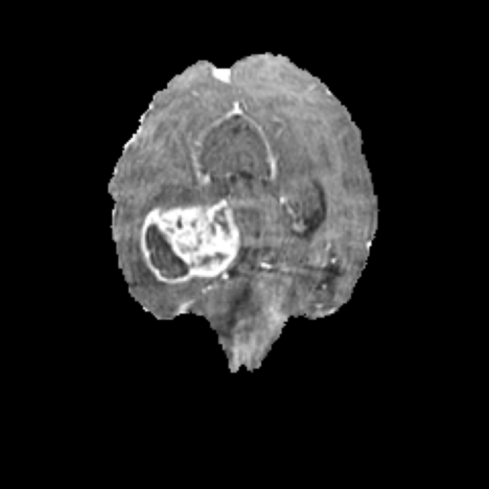
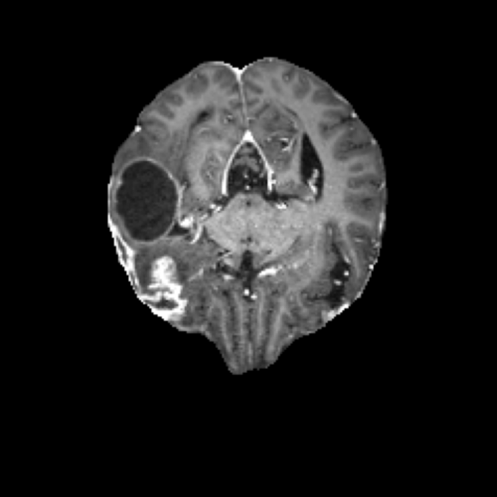
Unlike pre-operative imaging, where the tumor usually presents as a continuous mass, post-operative MRI scans are significantly more complex. Surgical intervention radically alters the brain anatomy by removing large portions of the tumor, which are replaced by a fluid-filled Resection Cavity (RC).
Furthermore, the surgical site often exhibits non-specific changes such as blood products, surgical debris, and inflammation. These artifacts can easily mimic the appearance of residual Enhancing Tumor (ET) on T1-weighted contrast-enhanced (T1c) sequences, while post-surgical edema can be mistaken for Surrounding Non-enhancing FLAIR Hyperintensity (SNFH). This dramatic anatomical shift makes automated segmentation of residual tumors a highly challenging task, prone to both false positives and missed recurrences if the model is not properly calibrated.
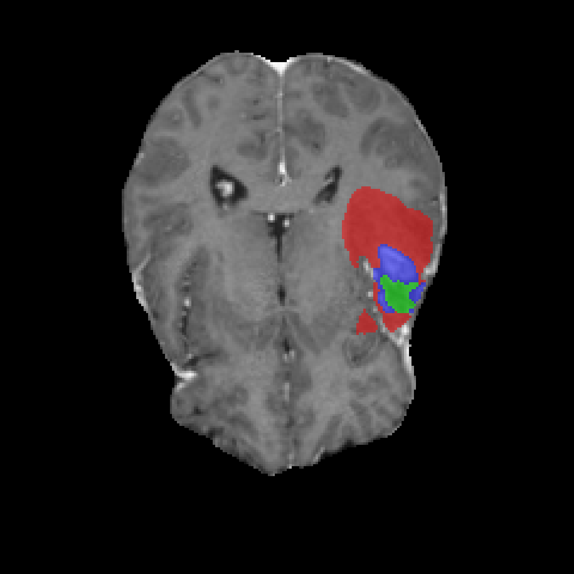
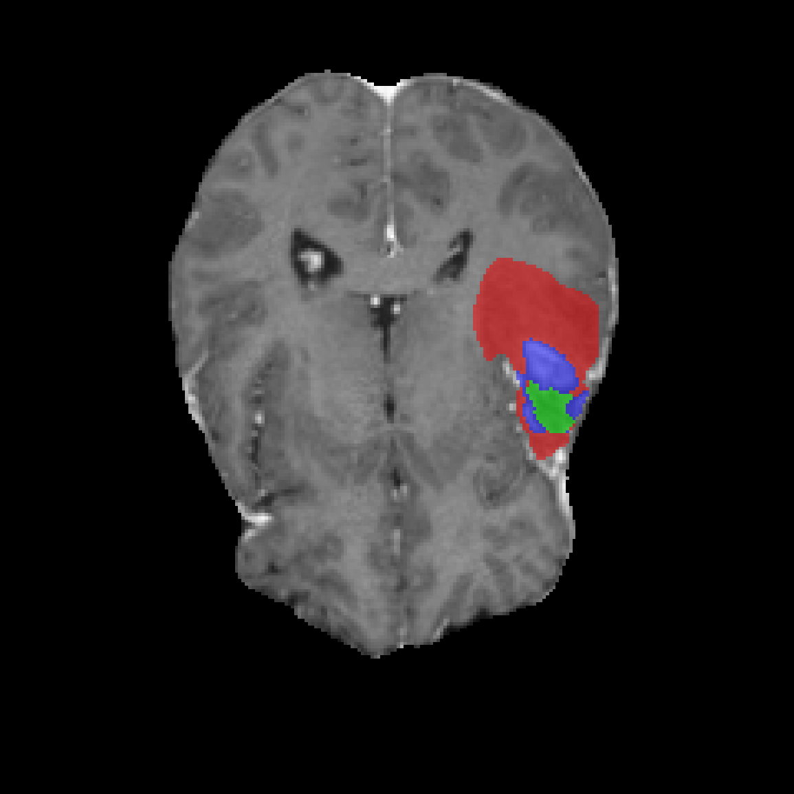
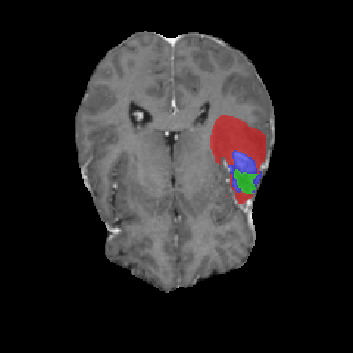
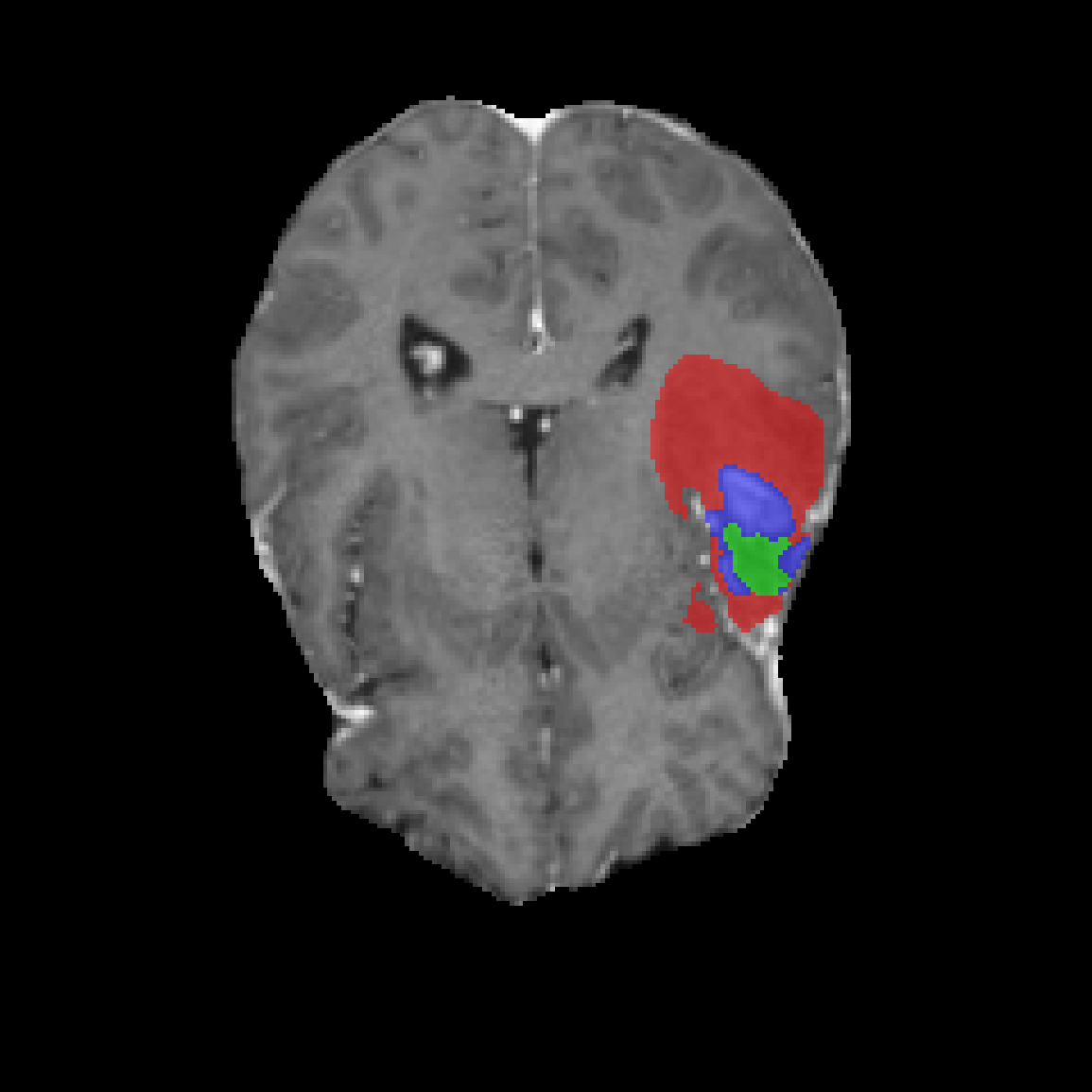
| T1c | GT | Baseline | Norm Variant | SACA Variant | |
|---|---|---|---|---|---|
| Case 1 | 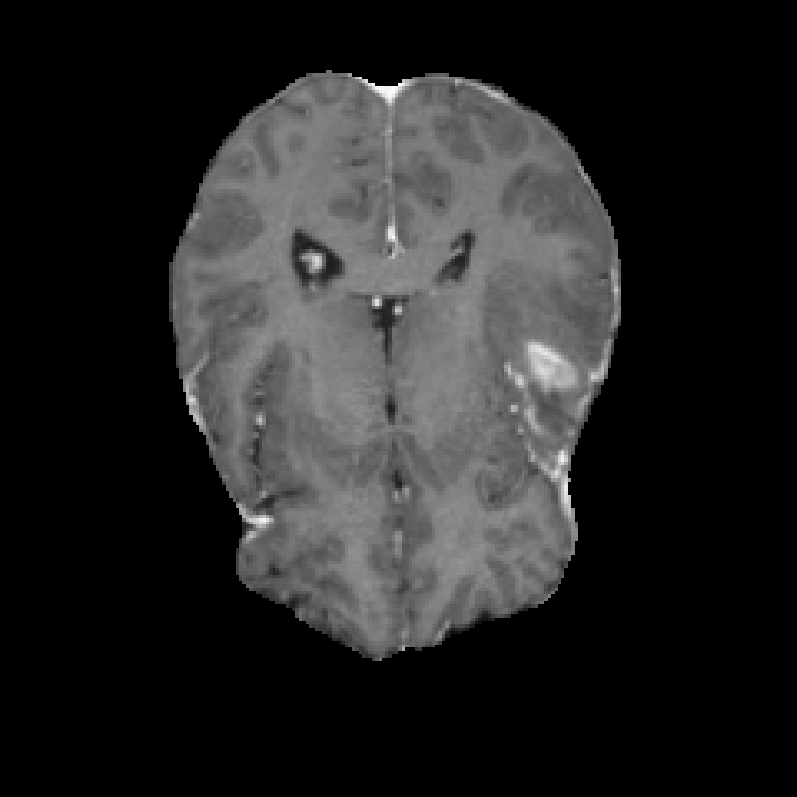 |
 |
 |
 |
 |
| Case 2 | 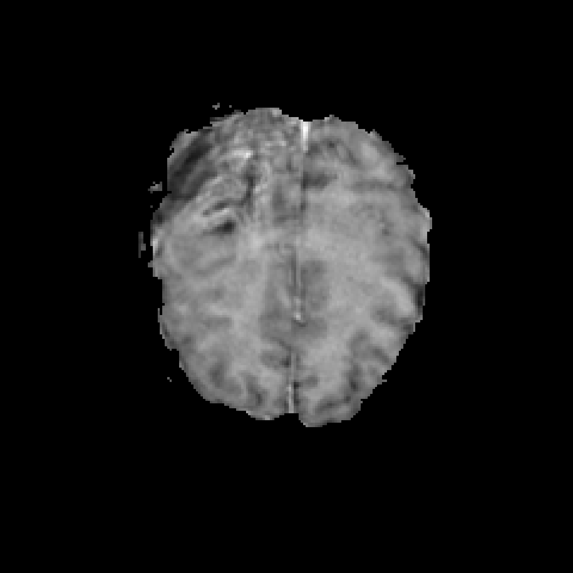 |
 |
 |
 |
 |
| Case 3 | 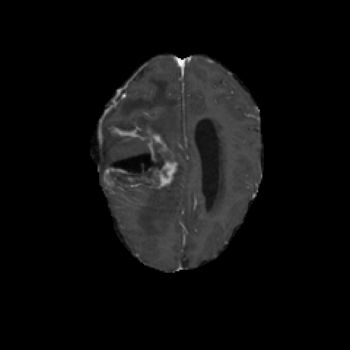 |
 |
 |
 |
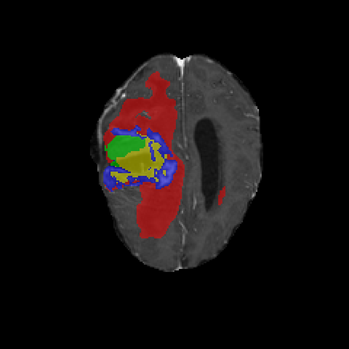 |
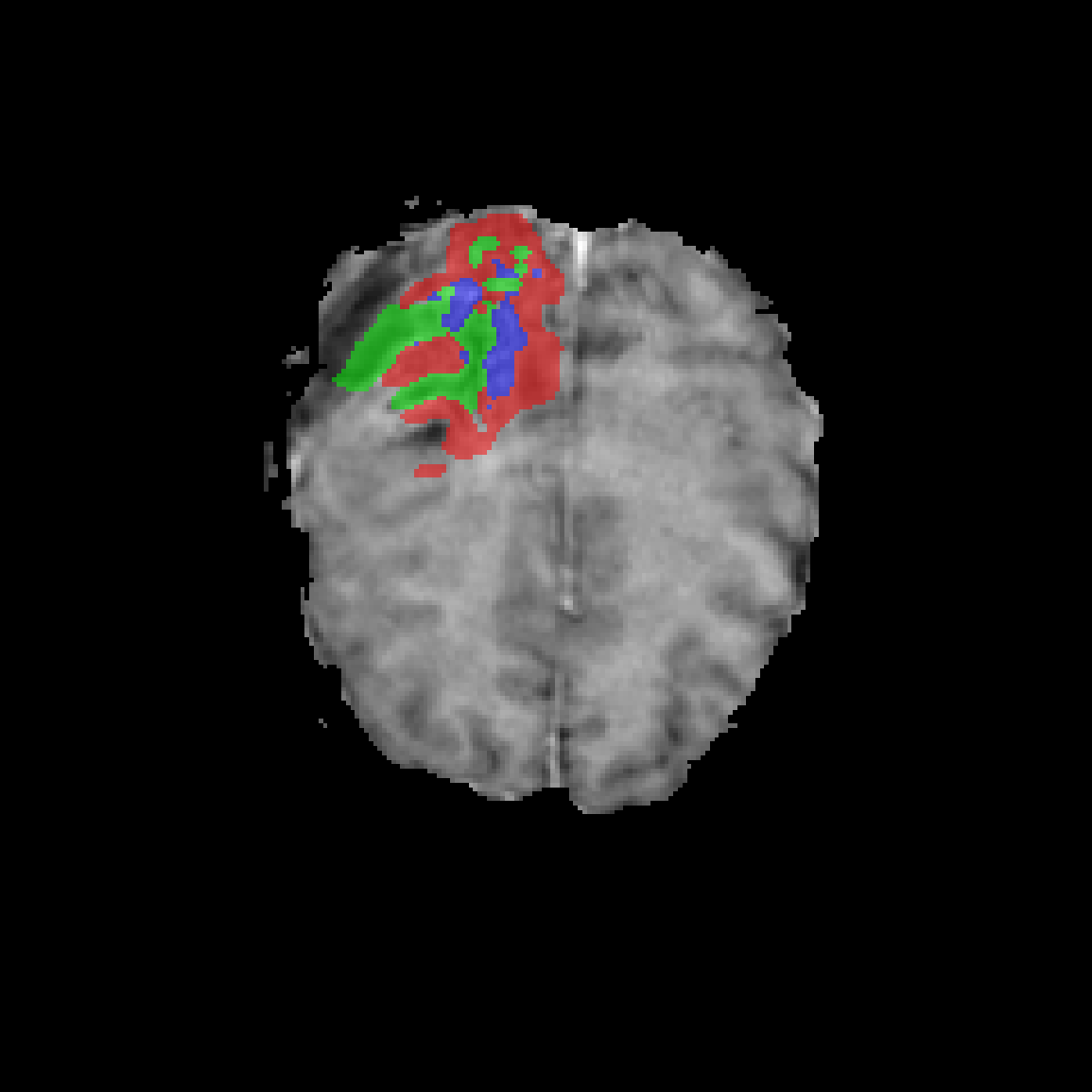
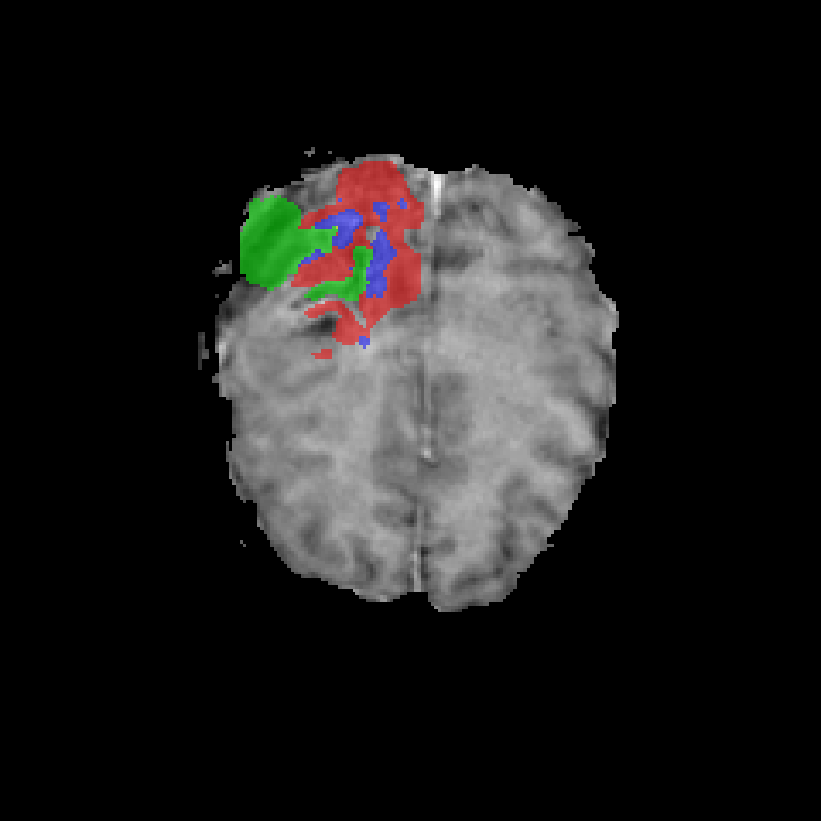
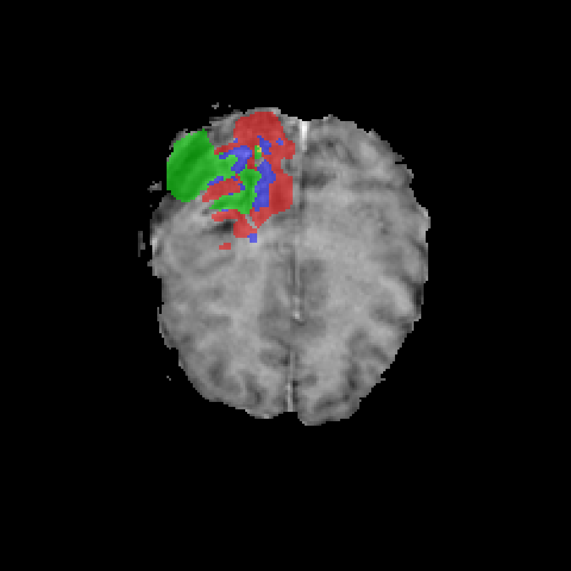
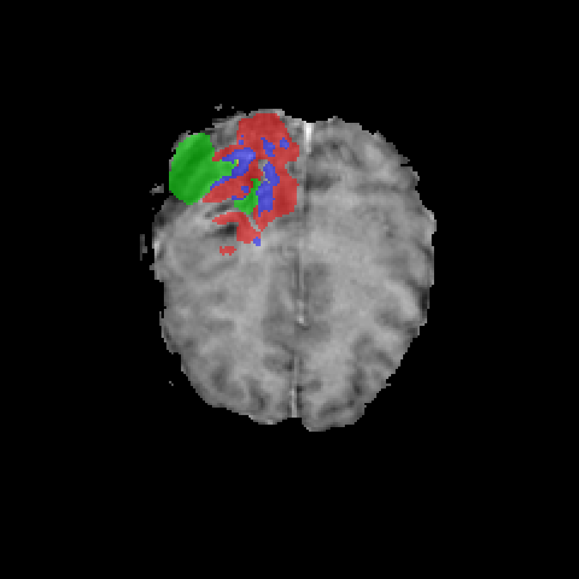
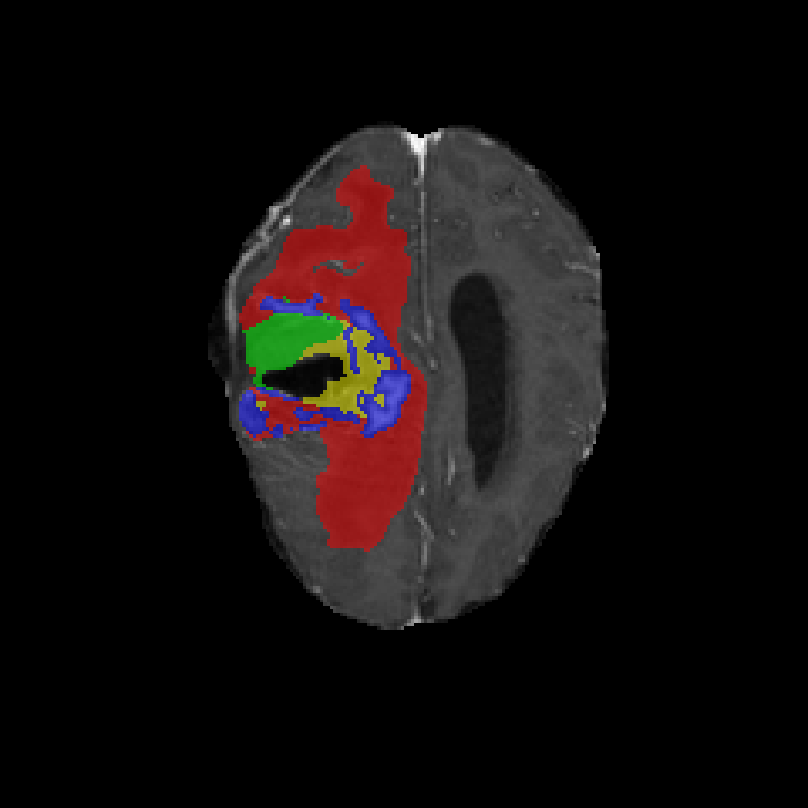
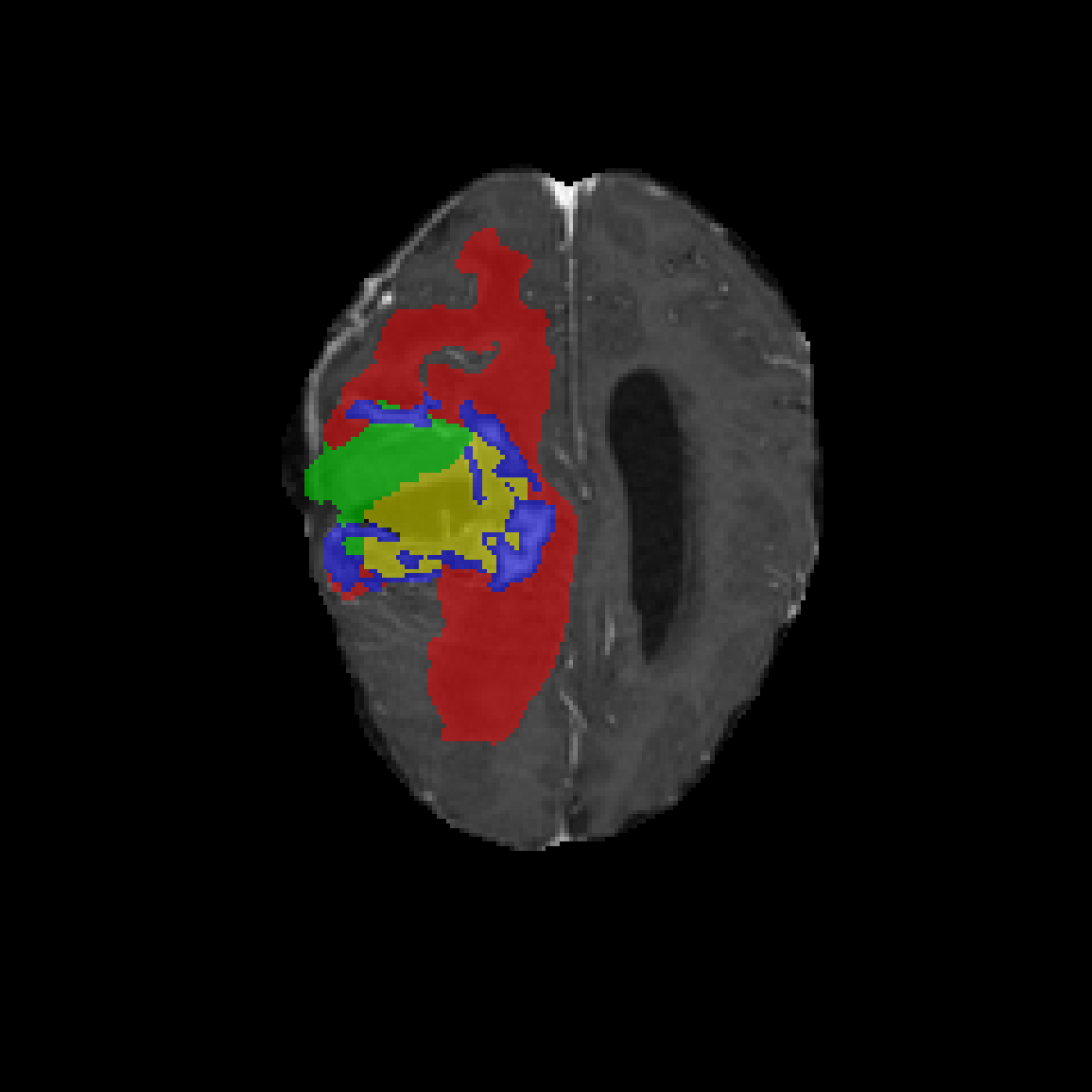
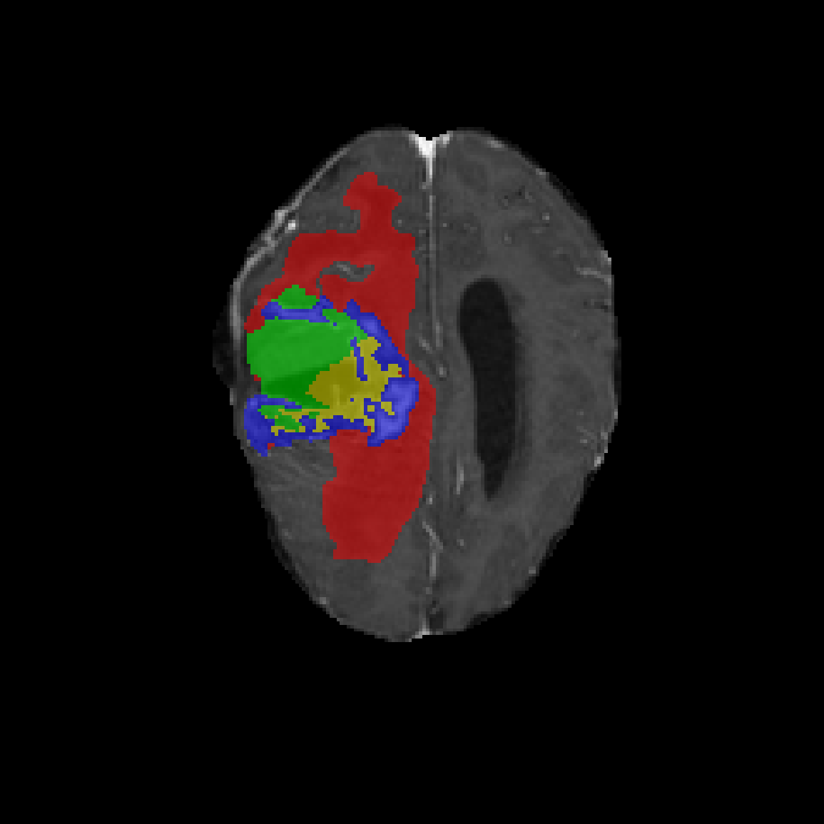
Qualitative results. Columns (left to right): T1c axial slice, Ground Truth (GT), Baseline, Norm Variant, and SACA Variant. Segmentation labels: ET (blue), TC (yellow), RC (green), and SNFH (red). Case 1: WL Dice > 0.95; Case 2: WL Dice ≈ 0.80; Case 3: morphology featuring a large resection cavity.
@misc{crişan2026postoperativegliomasegmentationloss,
      title={Post-Operative Glioma Segmentation via Loss Stabilization, Normalization and Subspace Attention},
      author={Alexandru Crişan and Diana Borza},
      year={2026},
      eprint={2607.22749},
      archivePrefix={arXiv},
      primaryClass={cs.CV},
      url={https://arxiv.org/abs/2607.22749},
      note={Accepted at ICANN 2026}
}